-
Industrial CT and X-ray
Inquiry
inspection systemMatsusada Precision offers a wide range of non-destructive X-ray inspection systems.
Engineered with our proprietary technology, these systems provide detailed internal visualization, optimized for electronic device applications. -

Benchtop X-ray Microscopy
Learn More
for NDT and CTTop-View Type Microfocus X-ray Inspection System
Benchtop with High Magnification
Compact and Powerful
Top-Class Image Resolution -

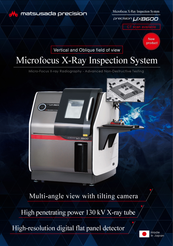
Microfocus X-ray
Learn More
Inspection SystemSupports top-view and oblique-angle CT scanning
Multi-angle view with tilting camera
High penetrating power 130 kV X-ray tube
High-resolution digital flat panel detector -
Microfocus
Learn More
X-ray Inspection
for NDT and CTTop-view type floor standing X-ray system
Top-Class Image Resolution
Small footprint X-ray system with precise and large scanning
Latest 3D analysis algorithm
Benchtop X-ray inspection
(Top-view type)
-
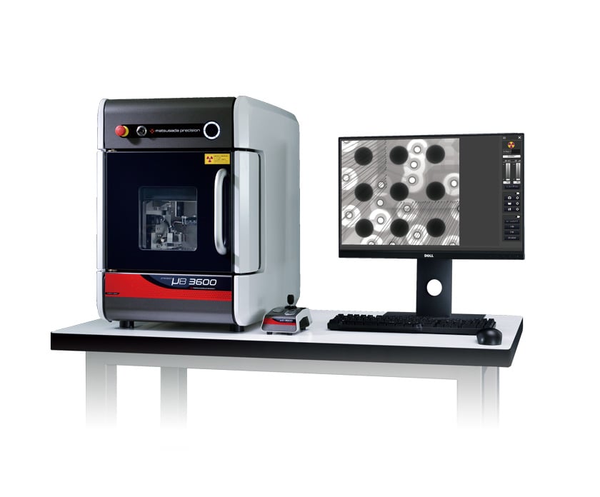
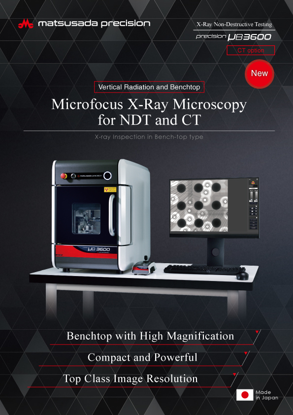
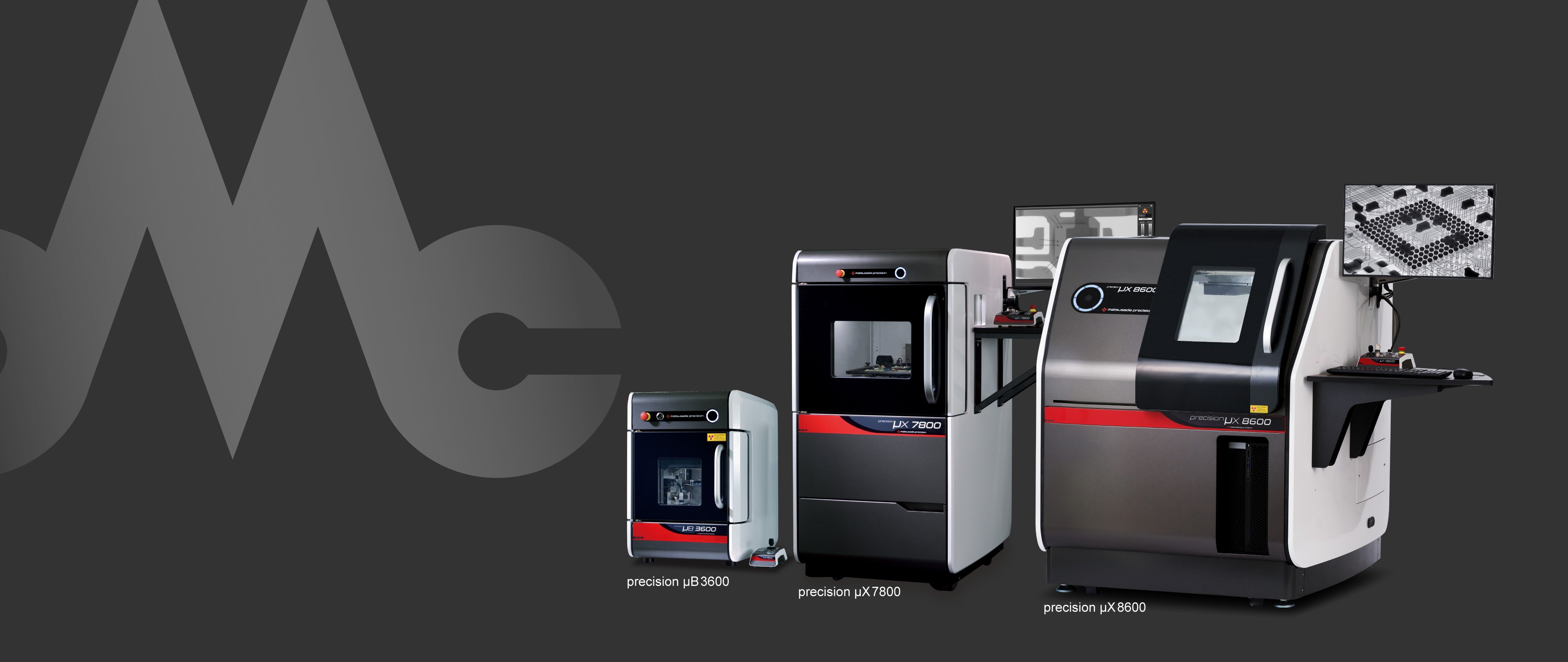
precision µB3600
- X-ray tube voltage
- 90 kV
- Minimum focal diameter
- 5 µm
Benchtop High Definition- Benchtop with High Magnification
- Compact and Powerful
- High-Resolution Digital Flat Panel Detector
- Sample table size X: 120 mm x Y: 140 mm
-
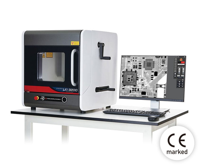
precision µB3200
- X-ray tube voltage
- 60 kV
- Minimum focal diameter
- 5 µm
Benchtop Entry Model- Suitable for small circuit boards and small components
- The maximum monitor image magnification is 125 times
- Easy maintenance
- Sample table size X: 225 mm x Y: 280 mm
-
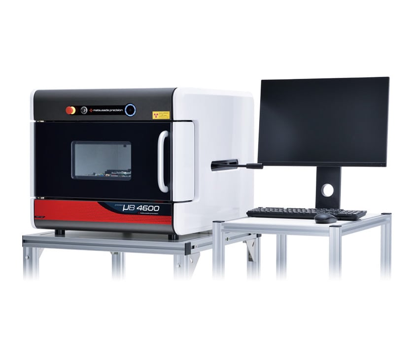
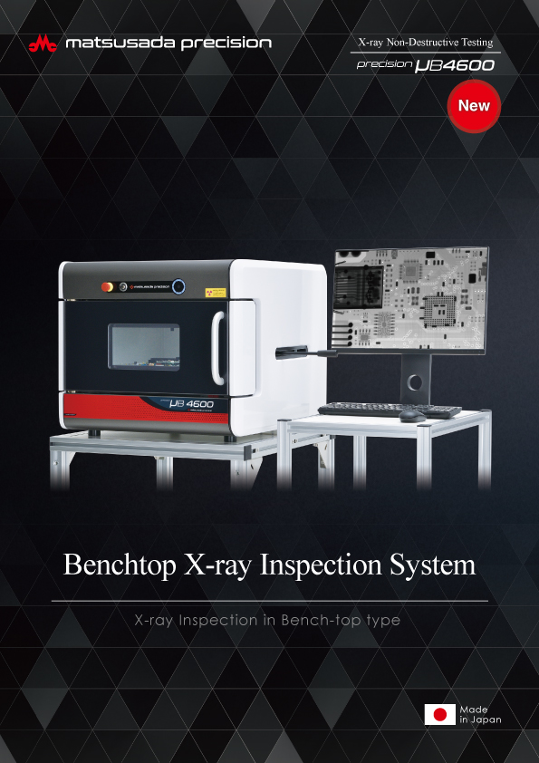
precision µB4600
- X-ray tube voltage
- 40 to 90 kV
- Minimum focal diameter
- 5 µm
Benchtop Manual Stage- Compact and Powerful
- Choice of X-ray sources and larger cameras
- Accommodates large samples for inspection
- Sample table size X: 330 mm x Y: 340 mm
X-ray Inspection Systems
(Top-view type)
-
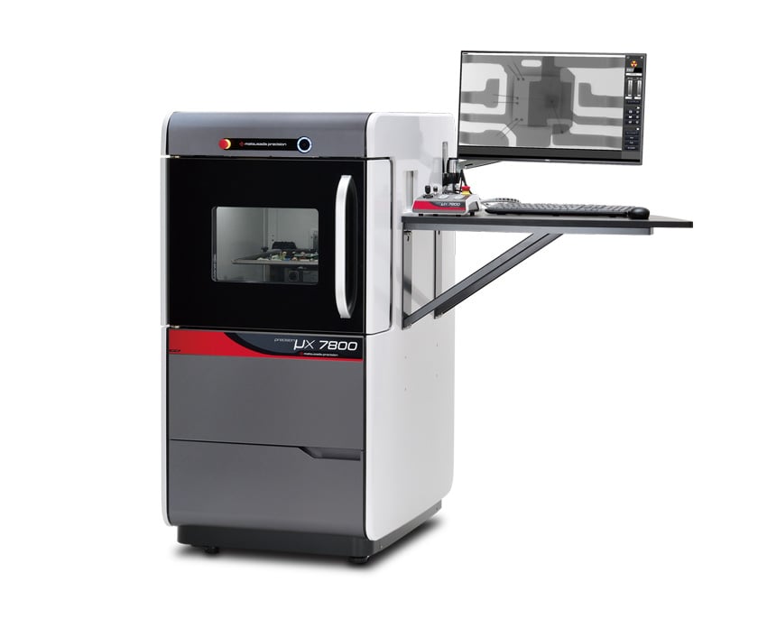
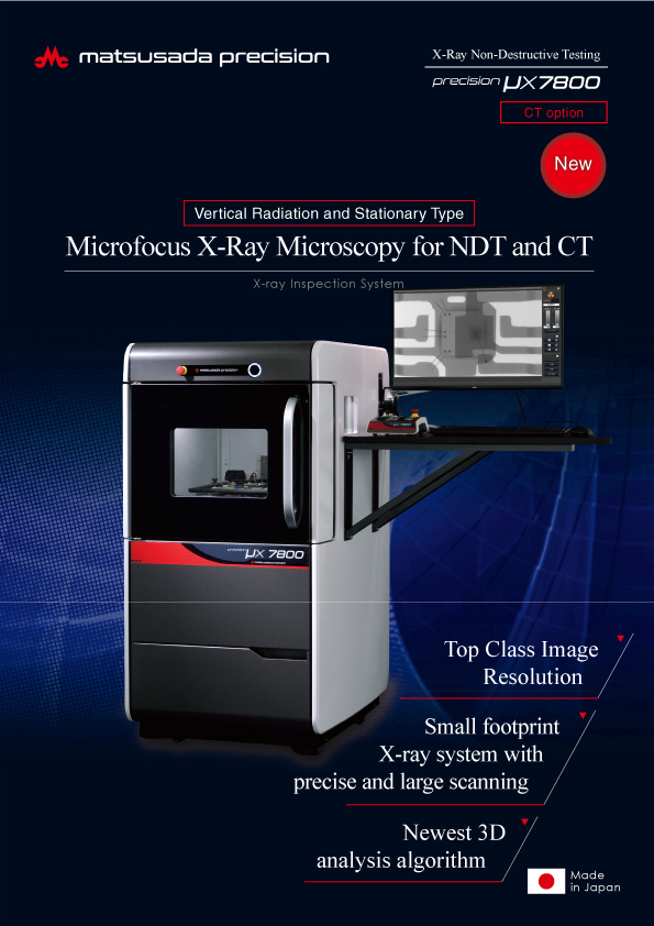
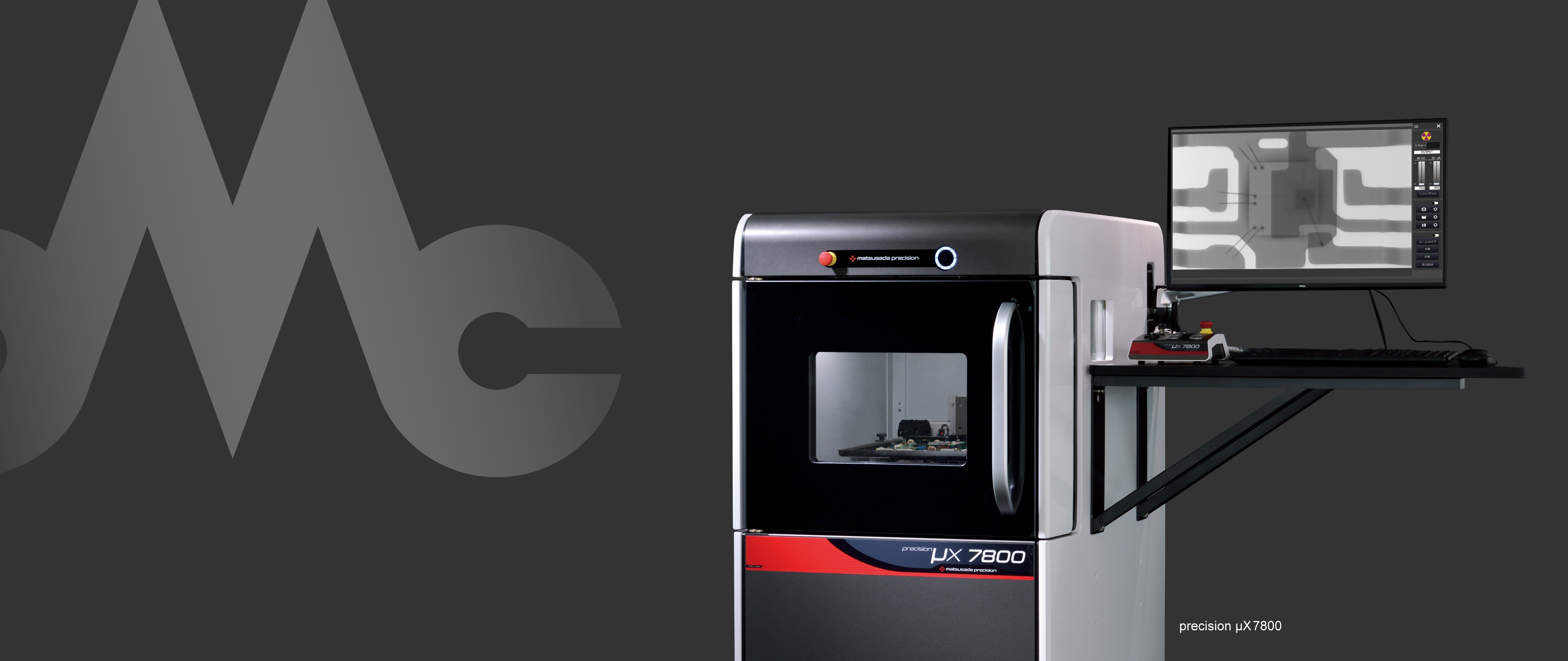
precision µX7800
- X-ray tube voltage
- 90 kV
- Minimum focal diameter
- 4 µm
Compact Standard Type- High-Resolution Digital Flat Panel Detector
- Compact but with a Wide Field of View and High Magnification
- State-of-the-Art 3D Analysis Algorithms
- Sample table size X: 265 mm x Y: 350 mm
-
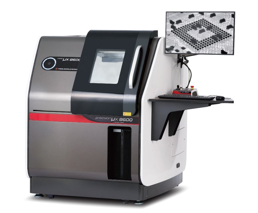
precision µX8600
- X-ray tube voltage
- 130 kV
- Minimum focal diameter
- 5 µm
For Printed Circuit Boards- Versatile Shooting with Tilting Camera
- High penetration power with 130 kV X-ray tube
- High-Resolution Digital Flat Panel Detector
- Sample table size X: 400 mm x Y: 450 mm
-
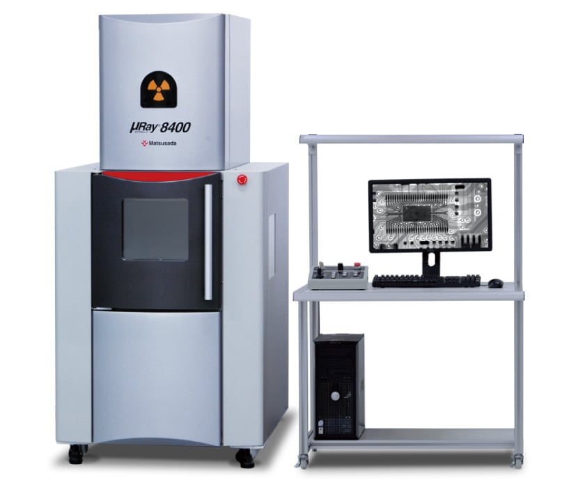
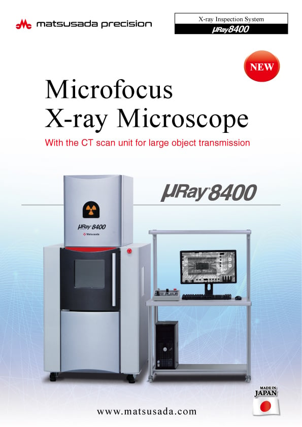
µRay8400-LF
- X-ray tube voltage
- 130 kV
- Minimum focal diameter
- 5 µm
Compact High-Power Type- High penetration power with 130 kV X-ray tube
- High-Resolution Digital Flat Panel Detector
- Sample table size X: 350 mm x Y: 450 mm
Micro-CT Scanners
(Side-view type)
-
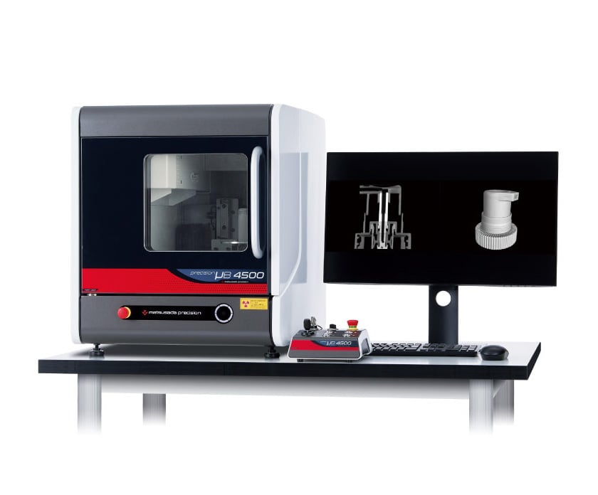
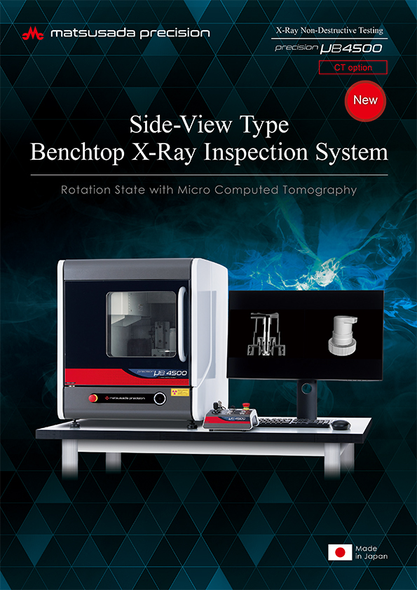
precision µB4500
- X-ray tube voltage
- 90 kV
- Minimum focal diameter
- 5 µm
Benchtop High Definition- Benchtop Size CT Imaging
- Compact and Powerful
- High-Resolution Digital Flat Panel Detector
- Sample table size Φ: 120 mm
-

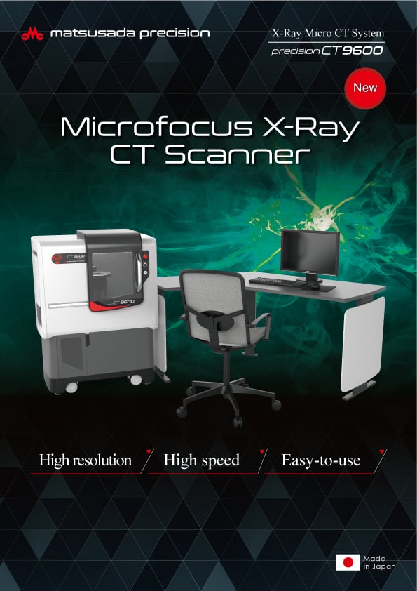
precision CT9600
- X-ray tube voltage
- 130 kV
- Minimum focal diameter
- 5 µm
Micro-CT Specific- High-Speed CT Imaging
- High-Resolution Digital Flat Panel Detector
- Intuitive UI Design
- Sample table size Φ: 200 mm
-
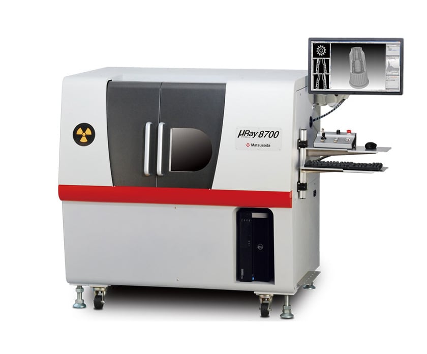
µRay8700/µRay8760
- X-ray tube voltage
- 130 kV
- Minimum focal diameter
- 5 µm
For Large Sample- Suitable for large sample inspection
- Compact but with a Wide Field of View and High Magnification
- Sample table size Φ: 200 mm x H: 340 mm
Features
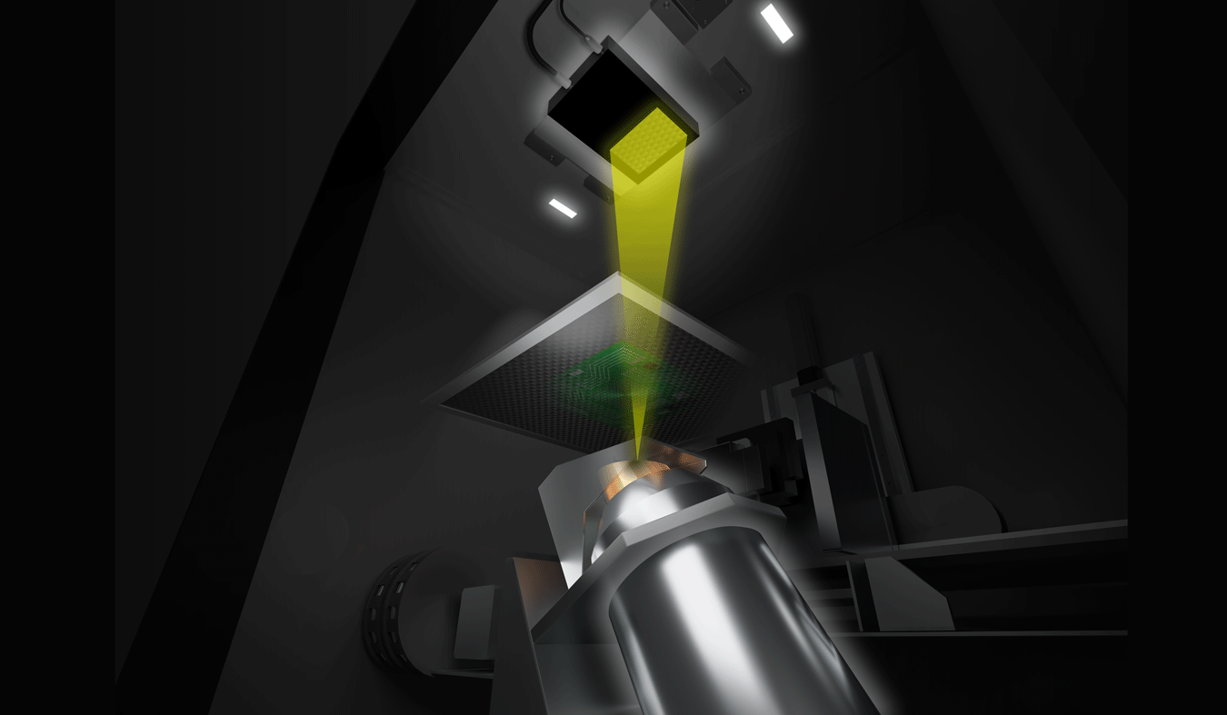
High-quality X-ray tube developed by Matsusada Precision
In order to produce clear X-ray images, the X-ray tube and high-voltage power supply have been independently developed to generate high-quality X-rays with low noise.
The focal spot size of the X-ray tube is the smallest in the industry for a sealed reflective type, and the low noise from the high-voltage power supply significantly reduces X-ray blurring.
New HDR-Pro image processing for high dynamic range radiography
Radiographic images produced with HDR-Pro feature wide dynamic ranges. The HDR-Pro ensures clear visualization, preserving details in both highlights and shadows.


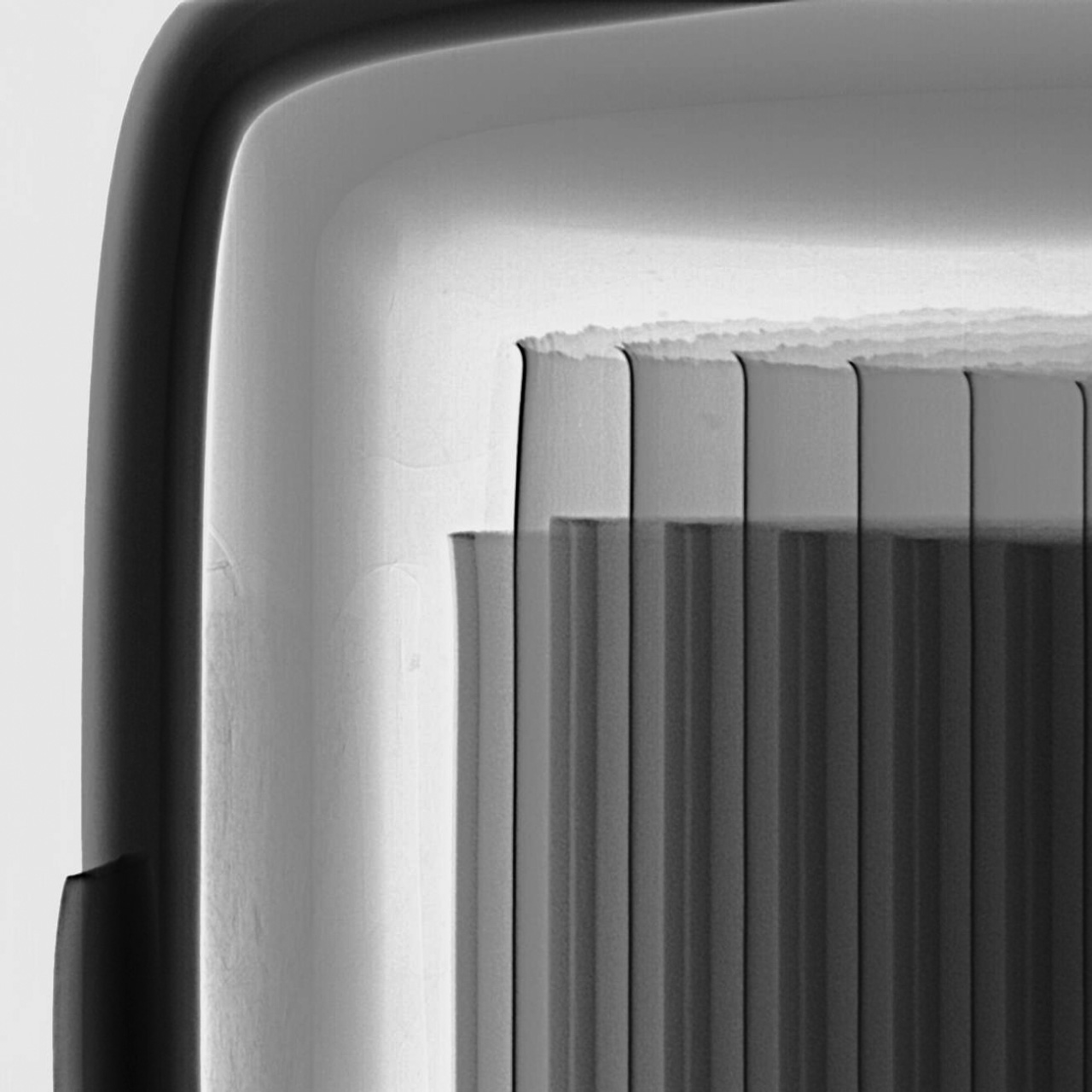
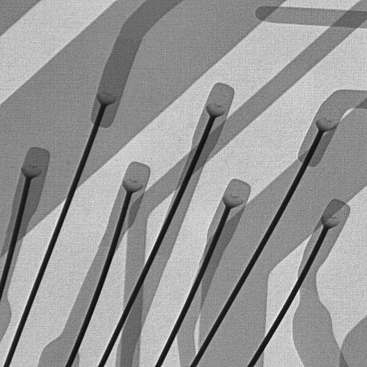
Image Gallery


Applications
-
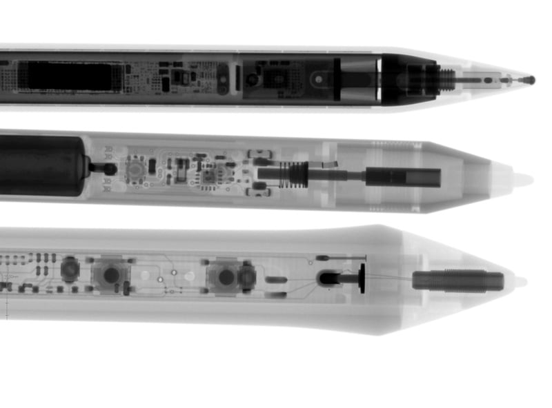
Apple Pencil vs. Surface Pen vs. Wacom Tablet Pen
The Apple Pencil, known for its sophisticated features, such as its tilt and pressure sensitivity that enables smooth writing...
See more -
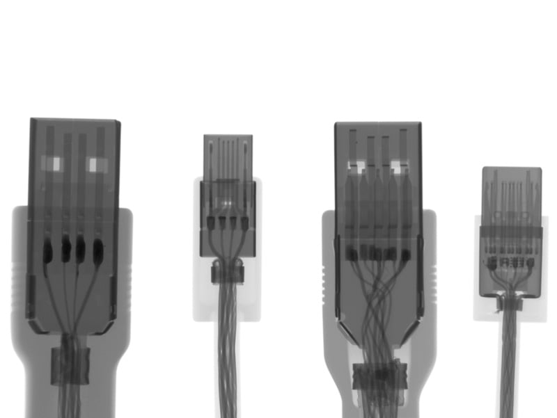
USB Cable Analysis
The X-ray images compare four different USB cables: USB 2.0, USB Micro, USB 3.0, and Type-C cables...
See more -

Various Battery Types
The X-ray comparison shows images of four different batteries: an AA-size manganese battery, an alkaline battery, a nickel-metal hydride battery...
See more -
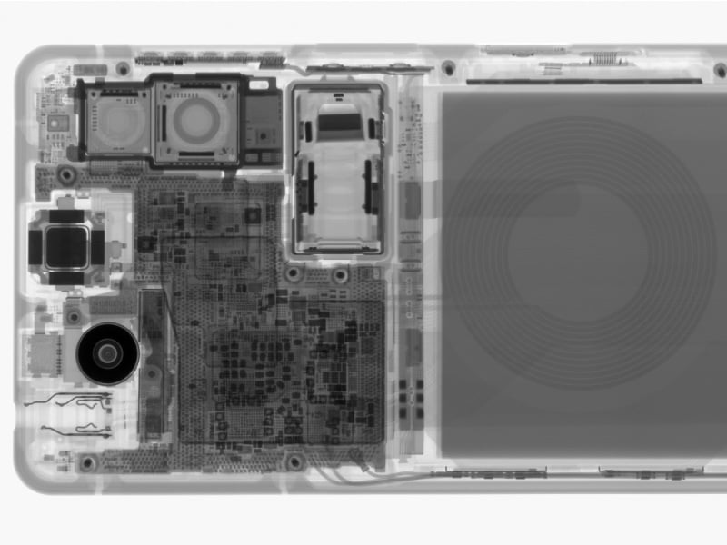
Wireless Charger vs. Smartphone
The X-ray images reveal the internal structures of both devices. Wireless charging operates without physical connections...
See more -
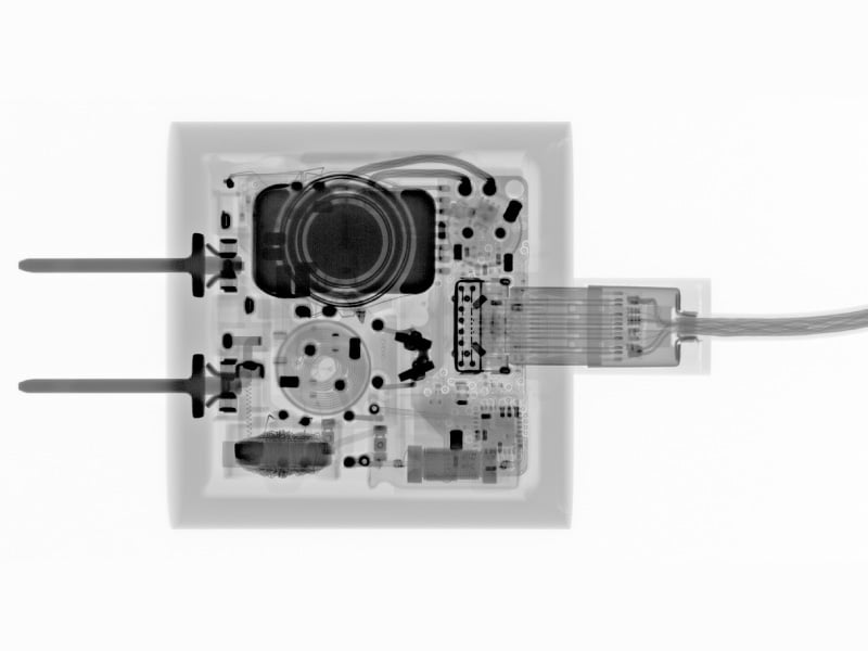
AC Adapter
The X-ray scan of Apple's genuine A1720 charger (18W, USB-PD-compatible) shows the circuit board, ...
See more -
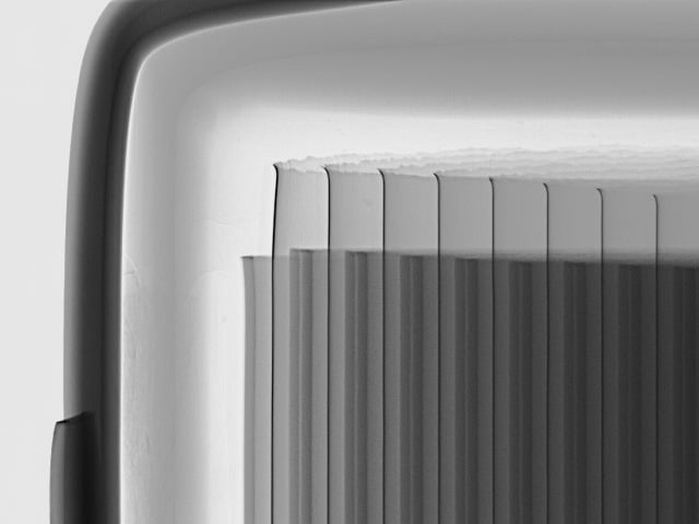
Lithium Ion Battery
Lithium-ion batteries (LIB, LFP, NMC) consist of alternating layers of cathode plates, anode plates, and separators inside a case. X-ray inspection systems are used to inspect the quality of lithium-ion batteries...
See more -
BGA Solder Joint
The causes of BGA malfunctions include bump voids, cracks, warpages, and deformations. BGA bumps are located on the underside of the chip, and we cannot visually inspect them...
See more